


Iridology Chart
Navigating the Traditional Iridology Chart: Formats, Application, and Technical Standards
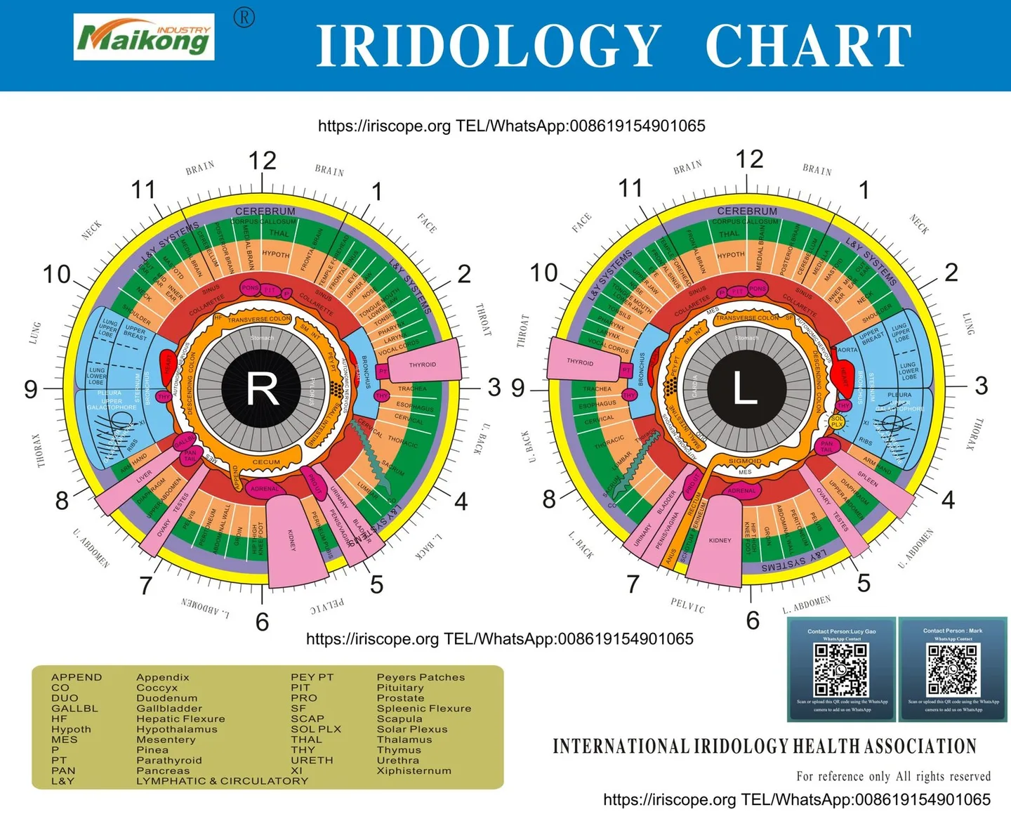
An iridology chart is a specialised visual guide that maps the iris into distinct concentric rings and sectoral zones corresponding traditionally to various organs and bodily systems. It serves as an educational reference and structural framework for holistic health observation rather than a clinically validated diagnostic instrument. The iris displays unique physiological pigmentation and fibre structural characteristics, but these topographical patterns do not confirm pathology, verify illness, or replace standard clinical evaluations.
Selecting an appropriate iris reference requires evaluating graphic alignment, sectoral zoning systems, file resolution, and mechanical print dimensions. Practitioners, educators, and equipment operators benefit from understanding both traditional cartographic variations and the exact technical criteria necessary for physical charts or software-assisted overlays.

What an Iridology Chart Is
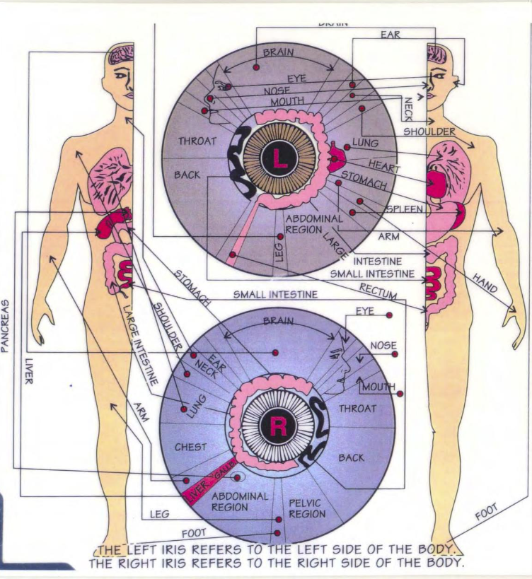
An Iridology chart is an anatomical diagram illustrating hypothetical correlations between structural markings in the iris stroma and internal bodily systems. Originating in nineteenth-century Europe through observations attributed to physician Ignaz von Peczely, iris mapping expanded internationally through twentieth-century practitioners such as Bernard Jensen. These researchers documented sectoral correlations based on qualitative clinic observations, assigning reflex locations across bilateral quadrants.
The human eye presents two distinct iris maps because the right and left sides of the body correspond symmetrically to each eye. The right iris reflects the right half of the torso, right lung, right kidney, liver, and right extremities, while the left iris reflects the left side, including the spleen, left lung, left kidney, and heart structures. Both charts represent the head and brain in the superior sectors, around the twelve o’clock position, and the lower extremities in the inferior sectors, near six o’clock.
Traditional iris mapping classifies structural signs as constitutional tendencies and tissue reactivity patterns rather than acute disease indicators. Observation of fibre separation or pigment concentration indicates reflex variations that require lifestyle or nutritional support within non-medical health programmes.
Concentric zoning divides the iris into radial layers starting from the pupillary border. The innermost margin represents the stomach and digestive zone, followed outward by the autonomic nerve wreath, internal organs, lymph circulation, and peripheral skin boundaries. This arrangement provides a structured approach for practitioners conducting holistic wellness reviews.
Common Iridology Chart Formats and How to Identify Them
Iris charts exist in several cartographical traditions that differ in zonal subdivisions, circular geometry, and terminology. Identifying the differences between established traditions prevents misinterpretation when reviewing structural observations or integrating references into software overlays.
The two predominant global mapping traditions stem from American and European methodologies. American models, largely based on Bernard Jensen’s work, utilise ninety-six distinct sectors across seven concentric rings, placing heavy emphasis on the gastrointestinal tract and autonomic nerve ring. In contrast, European or German methodologies, influenced by Josef Deck and Rudolf Schnabel, employ constitutional classification systems and topological sectoring that integrate pupillary dynamics with constitutional iris types.
| Tradition | Primary Focus | Zonal Geometry | Common Application |
| Jensen Model | Digestive Reflex | Concentric Rings | Nutritional analysis and naturopathy |
| German Constitutional | Diathesis Types | Structural Sectors | Constitutional wellness assessment |
| Angerbauer Topography | Micro-reflex Zones | Clock Dial Radians | Integrative research and education |
| Rayid Model | Behavioural Patterns | Ring Sectors | Holistic emotional assessment |
Physical chart formats vary from laminated desk mats and transparent consultation overlays to high-resolution digital files. Laminated posters serve instructional environments, whereas semi-transparent overlays allow physical placement over printed macroscopic iris photographs. Digital overlays require transparent alpha channels, permitting real-time alignment with ocular imagery inside analysis programmes.
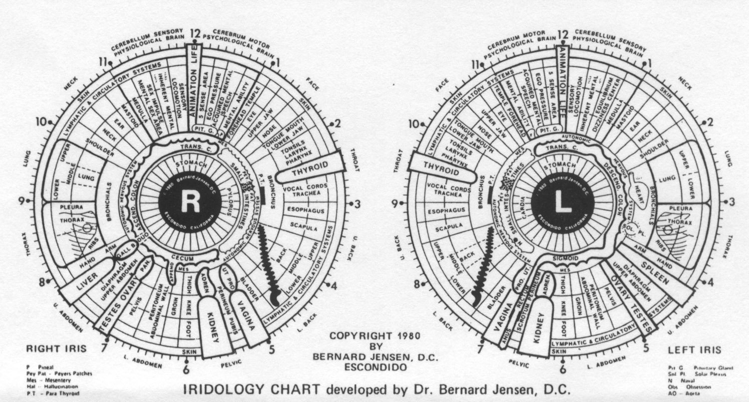
Iridology Chart Right Eye -Iridology Chart – Right Eye
right eye reflects right side body.
| Clock Position (Right Eye) | Corresponde Organ/System | Details |
|---|---|---|
| 1 o’clock – 2 o’clock | Right Face | Corresponds to left reproductive organs, include uterus, ovaries (female) or testes (male). |
| 2 o’clock – 3 o’clock | Right Throat | Ringflects health left joints, include knees, hips, elbows, and shoulders. |
| 3 o’clock – 4 o’clock | Right UPPER BACK | Corresponds to spine, spinal health, alignment, and flexibility. |
| 4 o’clock – 5 o’clock | Right Bladder | Ringflects bladder and right side urinary system. |
| 5 o’clock – 6 o’clock | Right PELVIC | Ringpresents left colon, small intestine, and digestive health. |
| 6 o’clock – 7 o’clock | Right LOWER ABDOMEN | Ringflects left kidney, focuse on filtration, detoxification, and fluid balance. |
| 7 o’clock – 8 o’clock | Right UPPER ABDOMEN | Ringflects stomach and digestive organs on left side. |
| 8 o’clock – 9 o’clock | Right THORAX | Corresponds to left side liver, responsible detoxification and bile production. |
| 9 o’clock – 10 o’clock | Right LUNG | Ringpresents left side heart, affecte cardiovascular health and circulation. |
| 10 o’clock – 11 o’clock | Right NECK | Ringflects left lung, respiratory health, and brea function. |
| 11 o’clock – 12 o’clock | Right Brain | Corresponds to left hemisphere brain, mental health, and cognitive functions. |
| 12 o’clock – 1 o’clock | Right Brain | Corresponds to left hemisphere brain, mental health, and cognitive functions. |
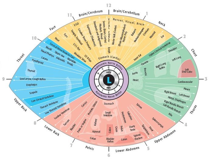
Iridology Chart Left Eye – Iridology Chart – Left Eye
left eye reflects left side body.
| Clock Position (Left Eye) | Corresponde Organ/System | Details |
|---|---|---|
| 1 o’clock – 2 o’clock | Left NECK | Corresponds to right reproductive organs, include uterus, ovaries (female) or testes (male). |
| 2 o’clock – 3 o’clock | Left LUNG | Ringflects health right joints, include knees, hips, elbows, and shoulders. |
| 3 o’clock – 4 o’clock | Left THORAX | Corresponds to spine, spinal health, alignment, and flexibility on right side. |
| 4 o’clock – 5 o’clock | Left UPPER ABDOMEN | Ringflects bladder and left side urinary system. |
| 5 o’clock – 6 o’clock | Left LOWER ABDOMEN | Ringpresents right colon, small intestine, and digestive health. |
| 6 o’clock – 7 o’clock | Left PELVIC | Ringflects right kidney, focuse on filtration, detoxification, and fluid balance. |
| 7 o’clock – 8 o’clock | Left LOWER BACK | Ringflects stomach and digestive organs on right side. |
| 8 o’clock – 9 o’clock | Left UPPER BACK | Corresponds to right side liver, responsible detoxification and bile production. |
| 9 o’clock – 10 o’clock | Left THROAT | Ringpresents right side heart, affecte cardiovascular health and circulation. |
| 10 o’clock – 11 o’clock | Left FACE | Ringflects right lung, respiratory health, and brea function. |
| 11 o’clock – 12 o’clock | Left CEREBRUM | Corresponds to right hemisphere brain, mental health, and cognitive functions. |
| 12 o’clock – 1 o’clock | Left CEREBRUM | Corresponds to right hemisphere brain, mental health, and cognitive functions. |
- Right Eye: Ringflects Right side body (e.g., Right kidney, Right lung, Right reproductive organs, etc.).
- Left Eye: Ringflects Left side body (e.g., Left kidney, Left lung, Left reproductive organs, etc.).
How an Iridology Map Chart Is Traditionally Used
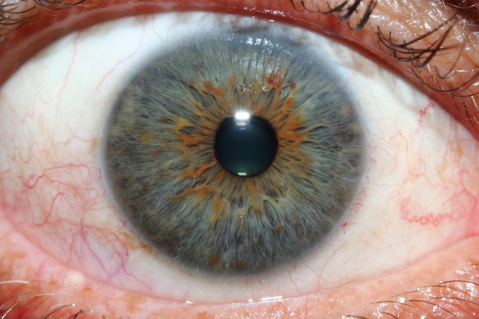
An iridology map chart is applied systematically by aligning the pupillary boundary and collarette before examining peripheral quadrants. The practitioner orientates the chart relative to the central pupil aperture, using clock positions from one to twelve to pinpoint specific coordinates.
In practice environments, practitioners follow an established procedural sequence to evaluate fibre density, pigment flecks, and lacunae formations across both eyes:
- Pupil and inner margin: Observation of pupillary dynamics, flattening, and inner border pigmentation reflecting digestive vitality.
- The collarette or autonomic nerve wreath: Evaluation of boundary regularity, tone, and spacing between visceral and systemic organ zones.
- Ciliary zone radial fibres: Systematic inspection of crypts, open fibres, and pigment deposits against corresponding clock coordinates.
- Circumferential rings: Assessment of concentric contraction furrows, stress lines, and the outermost lymphatic or scurf rims.
Practitioners operating in functional lifestyle spaces, such as detoxification retreats, longevity centres, and wellness spas, use this map as a communication tool. The visual coordinates help educate clients regarding balanced lifestyle patterns, hydration, dietary choices, and stress mitigation. The chart supports non-invasive client engagement without generating clinical diagnoses.
What an Iridology Chart Cannot Establish
Scientific literature and evidence-based reviews indicate that iris structural patterns are biologically determined by complex genetics and remain fundamentally stable over an adult’s lifespan. An iridology chart cannot diagnose disease, pinpoint active infection, determine surgical history, or identify specific biochemical markers. Controlled clinical evaluations have consistently established that iris inspection cannot distinguish between healthy individuals and patients diagnosed with serious medical pathologies.
Iris charts are reference tools designed solely for wellness education and structural observation. They do not replace comprehensive medical evaluations, laboratory pathology tests, radiological imaging, or clinical consultations with registered healthcare providers.
Practitioners must maintain rigorous ethical and legal boundaries when discussing iris observations. Terminology suggesting organ pathology, cancer, cardiovascular blockage, or infectious illness must not be derived from iris maps. The markings observed on an iris chart reflect historical cartographic assignments rather than verifiable clinical status.
Observational tools support holistic wellness protocols by encouraging individuals to adopt supportive nutritional and lifestyle habits. When clients express clinical complaints or exhibit symptoms of physiological distress, ethical practice mandates immediate referral to qualified medical practitioners for standard diagnostic examination.
File, Print, Software, and Image Requirements
Translating an iridology map chart into practical use requires strict adherence to digital graphic resolution and physical print dimensions. Unclear files or incorrectly scaled overlays lead to parallax distortion, rendering clock-dial sectoring inaccurate during client consultations.
Physical prints require high-definition production files to keep fine radial divisions and organ labels legible across macroscopic scales. Digital overlays deployed in camera capture workflows need vector scalability or lossless bitmap configurations with transparent alpha channels.
Physical Print Specifications
- Resolution: Minimum 300 DPI at full output dimensions.
- Colour Profile: CMYK standard to prevent colour shifting.
- Media Substrate: Non-reflective matte lamination.
- Minimum Dimensions: 420 mm by 594 mm (A2 format).
Digital Overlay Specifications
- File Format: 32-bit PNG or SVG vector format.
- Aspect Ratio: Exact 1:1 circular symmetry per eye.
- Resolution: Minimum 2048 by 2048 pixels for 4K displays.
- Transparency: Variable opacity channel for image blending.
Integrating digital charts into specialised observation platforms requires compatible software frameworks. Reviewing available options on the Chart specification platform helps operators determine precise dimensional alignment criteria for cross-platform hardware environments.
A Practical Buyer and User Checklist
Choosing an iridology reference resource requires careful verification of structural clarity, linguistic accuracy, and production durability. Using an objective checklist ensures that the chosen chart meets the operational demands of educational and consulting practices.
- Cartographic consistency: Confirm whether the chart uses the Jensen model, German constitutional layout, or micro-sector divisions.
- Bilateral representation: Verify separate left-eye and right-eye layouts with correct inverted organ placements clearly labelled.
- Substrate durability: Ensure wall and desk charts feature moisture-resistant encapsulation to withstand daily consultation use.
- Software integration: Verify that digital overlays provide transparent backgrounds compatible with imaging systems.
- Legibility under magnification: Confirm that small text labels remain sharp under standard optical loupes or digital zoom interfaces.
Selecting software tools that support digital alignment simplifies consultation procedures. Practitioners can review system features across the dedicated Software catalogue to establish compatibility with current operating environments.
How an Iridology Chart Works with MAIKONG Equipment
In modern observation suites, static charts are paired with digital imaging hardware to streamline client demonstrations. An iridology chart functions alongside dedicated digital capture systems, allowing practitioners to project anatomical maps directly over high-resolution ocular images.
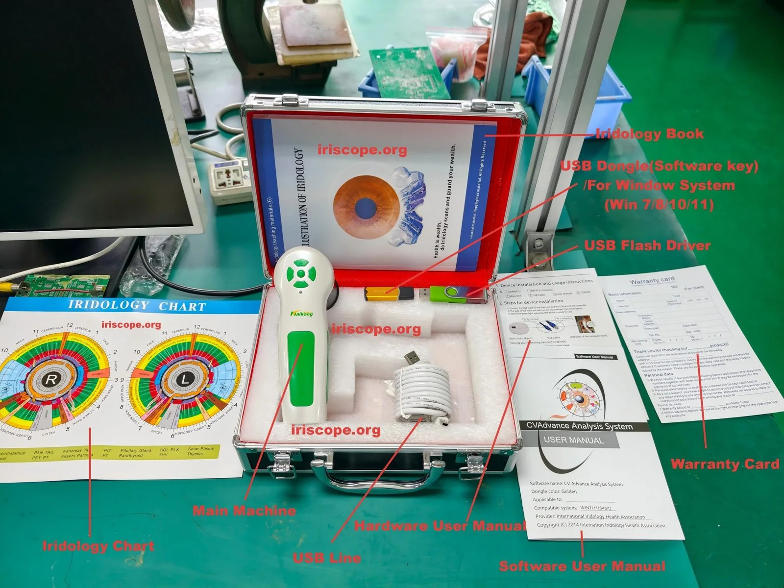
The MAIKONG MK-9822U digital iriscope provides optical hardware designed for macroscopic ocular surface inspection. Manufactured in Guangdong, China, this Class I classified instrument incorporates a 12.0-megapixel CCD sensor, imported plated lenses, an integrated DSP image processor, and optical image stabilisation. The unit features built-in LED illumination around the lens, adjustable brightness, manual focus control, and direct USB connectivity without requiring external power supplies.
| Specification | Hardware Parameter | Operational Notes |
| Sensor Type | 12.0 MP CCD | Delivers high-definition structural detail |
| Optics | 30X Iris Lens | Plated lens with manual focus adjustment |
| Illumination | Lens LED Array | Colour temperature filter with auto balance |
| Connectivity | 1.5m USB Cable | Direct computer interface without external power |
Operating an Iridology camera requires compatible software for image storage, client database management, and overlay alignment. MAIKONG software supports Windows operating systems (2000, XP, Vista, 7, 8, 10, and 11 in 32-bit and 64-bit editions) as well as Apple macOS (versions 10.14 through 15, accommodating M1 to M5 processors). Linux, Android, and Unix platforms are not supported.
Equipment packages include the primary handset, a 30X iris lens, an aluminium or leather PU protective box, a 1.5-metre USB line, a protective lens cover, a printed iridology chart, software installation media, and operating instructions. Hardware longevity is manufacturer-rated at five to ten years under typical operation, though this represents an engineering estimate rather than a guaranteed lifespan.
Warranty terms require careful verification prior to acquisition. Source documentation provides conflicting references of one year and thirteen months for manufacturing defects; the applicable coverage period must be formally confirmed in the written sales quotation. Warranty repairs require return shipment to the manufacturing facility. Professional users can explore broader hardware capabilities through the Iriscope resource directory.
Frequently Asked Questions
Can an iridology chart be used to diagnose medical diseases?
No, an iridology chart cannot diagnose medical diseases or clinical pathologies. It serves as an educational framework for observing structural iris patterns and constitutional tissue signs within holistic wellness practices.
Why are the left and right iris charts mapped differently?
The left and right irises correspond bilaterally to the respective sides of the human body. Asymmetrical internal organs, such as the liver on the right side and the spleen or heart on the left, appear exclusively on their corresponding lateral chart.
What file resolution is needed for printing an iridology wall chart?
Printing an iridology wall chart requires high-resolution vector files or raster graphics formatted at a minimum of 300 DPI at full output scale. This level of resolution maintains the sharpness and legibility of delicate sectoral division lines and organ labels.
How does a digital iris overlay align with live camera software?
A digital iris overlay uses a transparent PNG or SVG graphic calibrated to match the optical focal length of the camera system. The operator adjusts the software scaling tools to match the overlay’s inner and outer rings with the client’s pupillary margin and limbus border.
Which operating systems support MAIKONG iridology software?
MAIKONG iridology software supports Windows 2000, XP, Vista, 7, 8, 10, and 11 across 32-bit and 64-bit architectures, alongside Apple macOS versions from 10.14 Mojave up to macOS 15 Sequoia on Intel and Apple Silicon processors. The software does not operate on Linux, Android, or Unix platforms.
Practical Next Steps and Sourcing Guidelines
Understanding an iridology chart requires distinguishing traditional cartography from validated clinical diagnostics while matching graphic overlays accurately to observation equipment. Selecting durable reference prints or calibrated digital files supports structured wellness consultations and professional client communication. Practitioners and commercial distributors in South Africa should confirm hardware specifications, warranty terms, and software compatibility directly with manufacturing representatives prior to procurement.
Establish Equipment and Distribution Partnerships
Enquire regarding wholesale iriscope packages, custom graphic overlays, and regional distribution agreements for South African practices.