Iridology Map
Iridology Map: Formats, Uses, Limitations and File Requirements
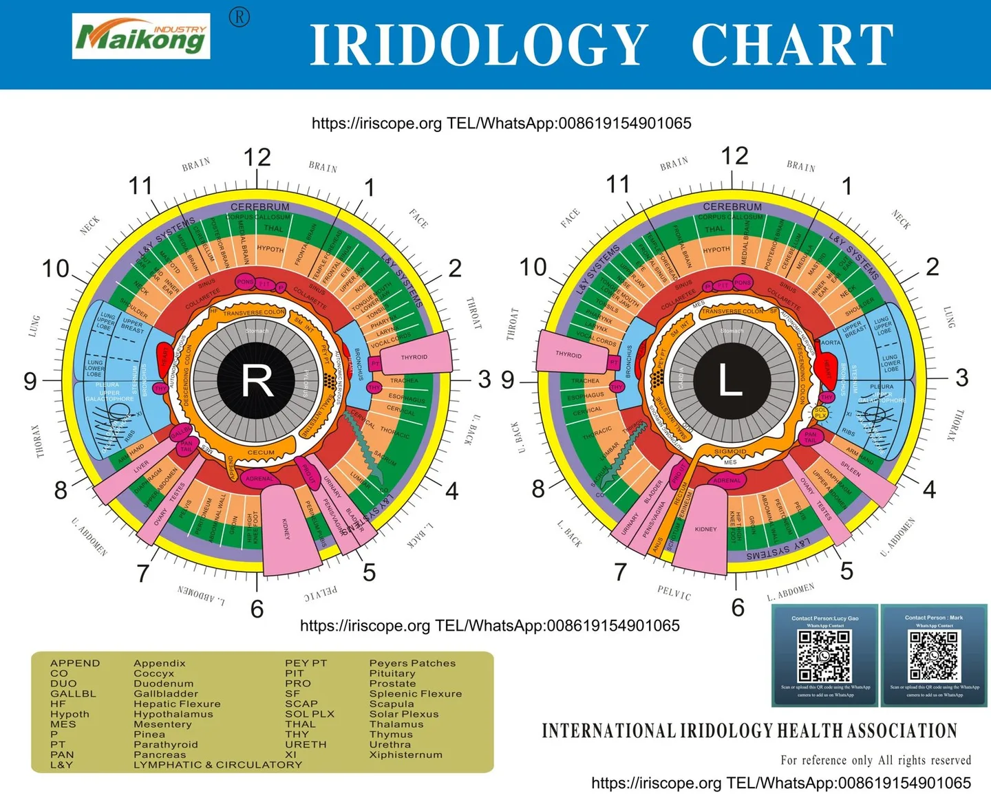
An iridology map is an illustrative reference diagram that divides the human iris into concentric rings and radial sectors associated with various body areas and physiological systems. Traditional practitioners use these illustrations during non-invasive client reviews to observe structural variations, pigment patterns, and iris fibre density. Such diagrams function strictly as educational frameworks and personal lifestyle assessment aids rather than instruments for medical pathology detection.
Selecting an appropriate reference chart depends on the display setting, the required level of anatomical sector detail, and the technical format needed for paper or screen examination. Comparing the operational scope, digital resolution, and system compatibility of iris mapping resources prevents common assessment errors and maintains professional standards.

Iris Topography: What an Iridology Map Contains
A standard iridology map arranges anatomical correlations across circular zones moving outwards from the pupillary margin to the outer ciliary edge. The inner zones represent the digestive tract, including the stomach and intestines, while peripheral rings correspond to the circulatory system, lymphatic structures, muscular frameworks, and skin. Radial segments divide the circle into clock-like intervals, separating upper body areas like the head and brain from lower structures such as the pelvic organs, kidneys, and legs.
Charts separate the left and right eyes to match bilateral human anatomy. The right iris diagram reflects the right side of the body, including the liver, ascending colon, and right lung, whereas the left iris diagram correlates with the left side, including the spleen, descending colon, and heart. Central reflex rings, historically known as the autonomic nerve wreath or collarette, mark the primary boundary between nutritional absorption areas and systemic organ zones.
Iris diagrams map symmetrical organs across both eyes while placing unilateral structures exclusively on their corresponding anatomical side. Confirming left and right eye orientation remains the most critical step before assessing topographical markings.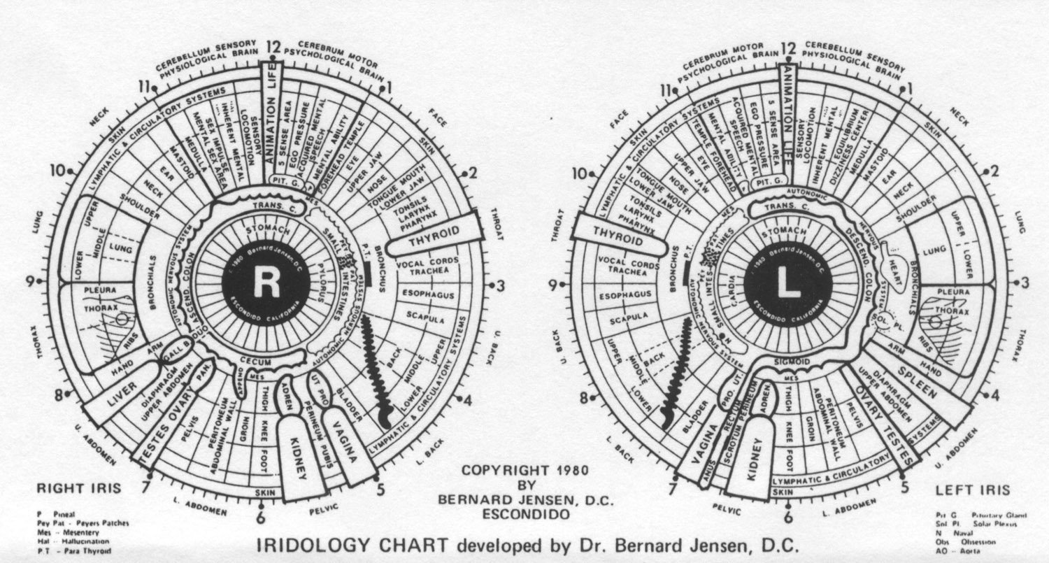
Traditional Formats for Iris Chart Resources
Iris reference diagrams are produced in several physical and digital layouts to accommodate distinct consultation environments. Large-format wall posters provide clear visual communication during client education sessions, while laminated desktop cards allow quick reference beside optical inspection devices. Transparent overlays and digital graphic assets can align directly with photographic images taken by an Iridology camera, assisting structured visual record-keeping.
Physical Chart Resources
- Rigid laminated desk reference cards
- Large wall-mounted educational displays
- Transparent plastic pupil-alignment overlays
- Printed multi-page sectoral reference manuals
Digital Chart Resources
- High-resolution scalable vector graphic files
- Transparent PNG overlays for photo software
- Integrated digital overlays in analysis programmes
- Printable PDF client report templates
Consultation spaces benefit from pairing a physical wall Iridology chart with matching digital assets inside viewing software. This balance enables practitioners to demonstrate general anatomical concepts on physical media while conducting high-magnification photograph reviews on a computer workstation.
Selecting a Map for Education or Wellness Practice
Evaluating an iridology map requires checking topographical clarity, sector labelling accuracy, and alignment with recognized historical traditions. Different historical approaches, including the European models stemming from Ignaz von Peczely and later American methods popularised by Bernard Jensen, feature minor variations in sector borders and terminology. Selecting a model with distinct boundary lines and standardised terminology reduces interpretive confusion across professional consultations.
Advantages
- Standardised sector boundaries simplify client communication.
- High-contrast radial markers accelerate image orientation.
- Complementary left and right templates reduce recording mistakes.
Limitations
- Minor sectoral variations exist between European and American schools.
- Crowded typography on small prints impairs legibility.
- Diagrams lack clinical capability to pinpoint active infections.
Practitioners working with dedicated Iridology Software often choose digital reference assets that match their software’s internal sectoral grid. Maintaining consistency between printed education sheets and digital overlay screens supports smooth client communication throughout health consultations.

File, Print and Digital-Use Requirements
High-quality digital iris charts require specific file characteristics to prevent pixel distortion and misaligned sector boundaries when enlarged. Raster images such as JPEG or PNG files must provide adequate pixel dimensions and colour contrast for printing or on-screen magnification. Vector formats like SVG or EPS allow scaling across large formats without loss of linework sharpness or font legibility.
- Resolution standards: Raster files should maintain three hundred dots per inch at intended print dimensions to preserve delicate zone labels.
- File architecture: Transparent PNG files allow clean positioning over raw photographic captures from a digital iriscope without obscuring underlying iris fibres.
- Colour balance: CMYK profiles prevent unexpected colour shifts on physical prints, whereas RGB profiles remain optimal for backlit monitor displays.
- Licencing terms: Verify commercial distribution rights for clinic report templates to avoid copyright infringements during public educational presentations.
When incorporating digital mapping files into client databases, uncompressed file formats prevent compression artefacts that might distort subtle boundary lines. Managing image files in structured folders categorised by client session date simplifies historical comparisons across successive wellness reviews.
A Practical Evaluation Checklist
Before purchasing or deploying an iridology map, practitioners should run a systematic review of the resource’s structural design and technical specifications. Ensuring proper anatomical labelling, clear ocular orientation, and suitable file formats prevents administrative delays in client management. This checklist assists wellness clinics, educators, and equipment distributors in selecting practical chart assets.
Chart Review Checklist: Confirm bilateral ocular labelling (Right/Left), verify pupil and collarette proportions, examine text legibility at arm’s length, check transparency settings on digital files, and inspect the required manufacturer software specifications.
Verifying that the resource includes clear non-diagnostic educational disclosures helps practitioners adhere to professional compliance standards. Pairing properly formatted templates with a compatible usb iriscope helps standardise photographic framing during routine wellness documentation.

Evidence, Limitations and Responsible Practice
An iridology map serves as an empirical, historical reference tool within complementary health and lifestyle education. Iris patterns reflect unique genetic tissue structures and phenotypic variations rather than real-time clinical biomarkers of specific disease states. Multiple scientific investigations have established that iris chart topography cannot detect internal malignancies, bacterial infections, cardiovascular blockages, or systemic pathologies.
Health consultations within any wellness clinic, natural health centre, or practice focusing on detox, ozone lifestyle support, anti-aging, or longevity must maintain precise boundaries. Content derived from iris charts is intended solely for personal wellness management, lifestyle habit coaching, and educational reference, and it cannot replace professional medical diagnosis, specialist examination, or clinical treatment. Practitioners must never offer medical confirmation, promise disease prevention, or claim that iris inspection substitutes for conventional pathology or general practitioner consultations.
Iris pattern reviews do not constitute medical examinations. When clients present with acute symptoms, severe pain, or persistent health complaints, immediate referral to qualified medical practitioners remains mandatory.
Responsible application of an Iriscope framework involves educating clients on general constitutional vitality, hydration habits, and nutritional balance. This disciplined scope fosters professional credibility and protects client well-being.
Hardware and Software Integration with Chart Resources
Modern digital wellness workflows combine high-definition optical sensors with dedicated software displaying dynamic chart overlays. Professional optical instruments capture fine stromal details, pupil dynamics, and radial fibres that correspond to physical reference cards. MAIKONG manufactures specialised optical hardware and software systems designed for international educational and lifestyle assessment facilities.
The MAIKONG Iris 12MP USB iriscope camera (Model MK-9822U) captures high-resolution imagery supported by a 12.0-megapixel CCD sensor, integrated LED illumination, and optical image stabilisation. The system connects directly via USB to desktop operating systems without external power cords, allowing operators to adjust focal clarity and LED brightness directly from the handset. Captured files integrate with proprietary analysis software that features client record databases, multi-language interfaces, and customizable reporting tools.
| System Type | Supported Versions | Operational Scope |
| Windows OS | Win 2000 to Win 11 | Supports both 32-bit and 64-bit system architectures |
| macOS | macOS 10.14 to 15 | Compatible with Apple Silicon M1 through M5 processors |
| Linux / Unix | Not Supported | Operating system environments currently lack software driver support |
Hardware longevity and manufacturer warranties represent important criteria for clinics and regional equipment distributors. MAIKONG provides professional Class I biochemical analysis category instruments manufactured in Guangdong, China, under standard ISO9000 factory management procedures.
| Item | Details | Terms Summary |
| Design Life | 5 to 10 Years | Typical service lifespan for electronic diagnostic accessories |
| Warranty Period | 13 Months | Coverage applies to manufacturing and material malfunctions |
| Warranty Service | Factory Return | Units are returned directly to the manufacturing facility for servicing |
The manufacturer offers customisation options including logo branding (minimum order: 50 units), graphic styling (50 units), and custom packaging (200 units). Clients and regional distributors should confirm applicable warranty periods and commercial supply terms in their official quotation.

Frequently Asked Questions
Is an iridology map a medical diagnostic tool?
An iridology map is not a validated medical diagnostic instrument and cannot identify or confirm medical conditions. It functions purely as an educational reference and lifestyle assessment tool within complementary wellness practices.
Can an iris map show internal organ diseases?
Scientific clinical research indicates that iris charts cannot detect organ pathology, infections, or systemic illnesses. Iris patterns reflect inherited structural tissue characteristics rather than active internal diseases.
Which computer operating systems support digital iridology charts?
Proprietary software suites support Windows XP, 7, 8, 10, and 11 across 32-bit and 64-bit systems, as well as modern macOS versions. Android, Linux, and Unix operating environments are currently unsupported.
What is the difference between right and left iris maps?
Right and left iris diagrams correspond to the bilateral anatomy of the human body. The right chart displays sectors for right-sided organs like the liver, while the left chart maps left-sided organs like the spleen and heart.
Practical Next Steps for Practice Resources and Distribution
Developing an effective wellness consultation environment requires selecting high-resolution iris reference maps that feature clear sector boundaries and responsible non-diagnostic disclaimers. Pairing durable physical charts with stable digital capture equipment establishes a reliable foundation for client education and constitutional wellness reviews. Practitioners and commercial business partners across South Africa can consult technical teams to determine compatible hardware, chart materials, and software solutions.
Equip Your Practice or Become a Regional Partner
Practitioners seeking professional hardware integration and businesses interested in South African regional distribution can connect directly with MAIKONG factory representatives for wholesale pricing, OEM branding, and technical support.
Contact US Online WHATSAPP: