


Basic Iridology
Basic Iridology: Principles, Observational Scope, and Boundaries
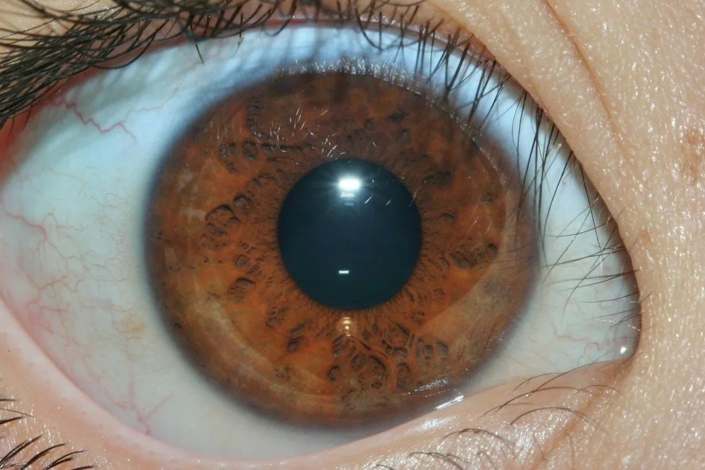
Basic iridology is the study and observation of visible markings, fibres, colours, and structural variations in the iris to explore potential constitutional tendencies. It serves as an educational framework within complementary health modalities, rather than a diagnostic procedure or medical assessment. Structural patterns in the eye reflect unique individual tissue arrangements that practitioners examine to discuss general wellness concepts.
Understanding where educational observation ends and clinical healthcare begins dictates the responsible application of this discipline. Clear boundaries, reliable reference charts, and objective imaging practices help beginners assess iris patterns without confusing exploratory observations with formal medical conclusions.
Defining the Scope of Basic Iridology
The core premise of basic iridology centres on reviewing the coloured part of the eye to gain insight into personal vitality, inherited strengths, and tissue integrity. Practitioners view the iris stroma as an intricate map where variations in pigmentation, shading, and fibre density correlate with conceptual bodily zones. This method provides educational insights into personal lifestyle habits, digestive balance, and nutritional tendencies.
Observation focuses on distinct morphological characteristics, including radial furrows, pigment spots, and concentric rings in the ciliary zone. An analysis of the eye provides a visual point of reference for holistic lifestyle evaluations rather than an inventory of disease states. It remains distinct from conventional ophthalmology, which assesses the eye for refractive errors, pathology, and structural optical disorders.
Basic iridology is an educational approach to observing and discussing visible iris features. It is not a medically established method for diagnosing disease, and an iris image cannot confirm a health condition. Anyone with symptoms or health concerns should consult an appropriately qualified healthcare professional.
Beginners learn to differentiate between innate iris colouration and acquired surface deposits without making therapeutic claims. By understanding that visible signs demonstrate physical constitution rather than acute medical issues, students maintain a disciplined, supportive approach to client well-being.

Iris Topography and the Role of the Iridology Chart
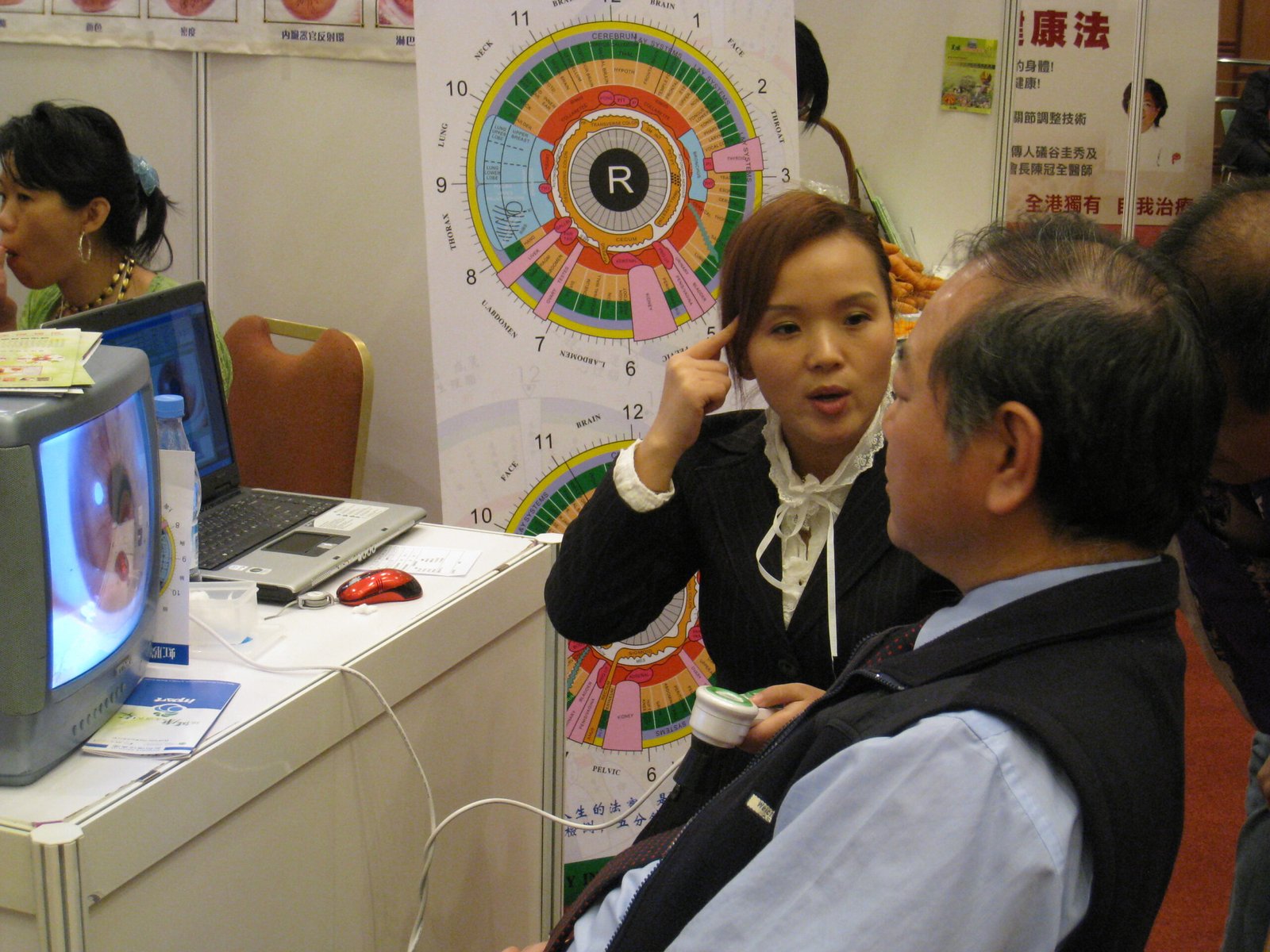
Topographical mapping forms the foundation of any observational study of the iris. Modern practitioners utilise a specialised Iridology chart that divides each eye into concentric rings and radial sectors corresponding to distinct functional areas of the body. The pupil margin represents the core digestive system, while outer concentric zones reflect systemic circulation, lymphatic activity, and the cutaneous border.
| Zone Layer | Relative Position | Conceptual Focus |
| Stomach Ring | Pupil Margin | Primary digestive uptake and mucosal tone. |
| Intestinal Band | Inner Ring | Nutrient assimilation and gut transit dynamics. |
| Autonomic Collarette | Mid-Periphery | Autonomic nervous system balance and energy distribution. |
| Organ Sectors | Ciliary Body | Tissue vitality across liver, respiratory, and structural systems. |
| Skin Border | Outer Margin | Eliminative dynamics through skin and lymphatic pathways. |
Each sector corresponds to specific metabolic systems, allowing users to cross-reference visible fibres with lifestyle practices. When practitioners evaluate a classical Chart, they maintain an educational approach, noting areas that might benefit from dietary refinement, improved hydration, or supportive rest. This map organises observations systematically, helping observers avoid haphazard assumptions across complex surface patterns.
Distinguishing Iris Assessment from Medical Practice
A critical responsibility in basic iridology involves acknowledging scientific limits and legal boundaries. Iris patterns are not scientifically proven to detect, confirm, or track specific internal diseases or acute infections. Clinical studies indicate that iris structures remain largely stable genetic markers over a lifetime, meaning transient illnesses do not cause immediate changes in ocular pigment or fibre alignment.
All content concerning results interpretation, bodily analysis, wellness clinics, and health management is strictly for educational reference. Observations must never be presented as a medical diagnosis, clinical confirmation, or justification to alter prescribed treatments.
When wellness centres incorporate iris analysis alongside modalities such as ozone therapy, detox protocols, anti-aging discussions, or longevity consultations, practitioners clarify that ocular review provides supportive context only. In cases involving severe symptoms, persistent pain, or metabolic dysfunction, immediate referral to a registered medical doctor is necessary. Ethical practice relies on complete transparency regarding what an eye evaluation can and cannot achieve.
Practical Observation Tools and Equipment Boundaries
Accurate visual evaluation in basic iridology depends on suitable magnification and steady, balanced illumination. Beginners frequently start with simple optical loupes equipped with side-angled penlights to avoid reflective glare from the cornea. As technical requirements expand, practitioners adopt digital imaging systems, commonly called an Iriscope, to capture detailed records for longitudinal study and client discussion.
Handheld Magnifiers
Simple dual-light optical magnifiers provide immediate visual inspections without requiring computer connectivity or complex software setups.
Digital Capture Systems
Dedicated digital units connect via USB to capture high-resolution imagery, enabling side-by-side analysis and organised historical record storage.
According to manufacturer specifications, the MAIKONG MK-9822U Iridology camera provides a 12.0-megapixel CCD sensor, LED illumination surrounding the lens, and an optical image stabiliser. The manufacturer notes that this unit operates via direct computer USB connection without external power supplies, using an adjustable focus ring to capture clear stroma images. Such digital devices function purely as capture instruments to facilitate educational dialogue, without providing automated medical assessments.
Record Management and Software Compatibility
Systematic record management allows holistic health educators to catalogue iris imagery alongside client history, dietary logs, and wellness goals. Using dedicated Iridology Software simplifies image archiving, visual grid overlays, and comparative evaluations over multi-year wellness programmes. Beginners should select software platforms that offer stable data backup options and run smoothly on their operating system.
Standard Visual Archiving
- Manual file sorting across separate folders
- Requires separate charting software for notes
- Relies on manual backup to external drives
Dedicated Wellness Software
- Integrated client database with time-stamped images
- Built-in digital topographical grid overlays
- Direct export of educational client summaries
Manufacturer documentation indicates that MAIKONG iridology software supports Windows systems from XP through Windows 11 in both 32-bit and 64-bit formats, alongside Apple macOS versions. The manufacturer states that the software platform includes multi-language options such as English, Spanish, French, and German, though Linux and Android environments are not supported. Operators should verify computer specifications and software licensing dongle requirements prior to implementation.


A Responsible Beginner Evaluation Checklist
Approaching basic iridology with appropriate care requires adhering to structured observational steps. Beginners must avoid rushing to conclusions based on isolated marks, focusing instead on overall constitution, symmetry between both eyes, and general tissue density. Evaluating the whole picture helps prevent speculative overinterpretation and supports a grounded educational conversation.
- Confirm ambient lighting is diffuse and position the client comfortably to minimise reflective glare on the cornea.
- Examine overall iris colouration to determine the foundational constitutional type: lymphatic, biliary, or haematogenic.
- Inspect the pupillary border to assess autonomic collarette tone and internal digestive ring alignment.
- Overlay a standard chart sector by sector to map visible crypts, radii, or pigment spots to conceptual organ zones.
- Document observations objectively without assigning disease names, using non-diagnostic terms focused on vitality and lifestyle balance.
Select Reliable Observation Equipment
Explore professional non-diagnostic imaging tools designed for clear ocular documentation and lifestyle tracking.
Frequently Asked Questions About Basic Iridology
Can basic iridology diagnose disease?
No, basic iridology cannot diagnose, treat, or detect specific diseases or medical conditions. It serves strictly as an educational observation tool to discuss tissue structure, constitutional tendencies, and lifestyle support. Individuals experiencing medical symptoms should always consult a registered physician for clinical diagnosis.
What does an iridology chart represent?
An iridology chart provides a conceptual map dividing the iris into concentric zones and sectors representing various bodily systems. It acts as an educational reference guide rather than an absolute anatomical guarantee. Observers use it to structure discussions around lifestyle, diet, and general vitality.
How does basic iridology differ from a medical eye examination?
A medical eye examination evaluates ocular health, visual acuity, and physical pathology using validated clinical diagnostic instruments. In contrast, basic iridology examines surface colours and fibre patterns purely for complementary health and lifestyle discussions. It does not assess refractive errors or identify clinical ocular diseases.
Do iris patterns change when a person recovers from an illness?
Scientific evidence indicates that the primary trabecular structure of the iris remains largely stable throughout adult life. Transient health fluctuations or recoveries do not produce immediate, dramatic structural reorganisations in the iris stroma. Claims of rapid ocular transformations following short-term health interventions lack robust scientific verification.
Summary of Educational Practice and Next Steps
Mastering basic iridology involves understanding the boundaries between constitutional exploration and clinical healthcare, while developing rigorous visual assessment skills. Relying on objective reference charts, maintaining high standards for photographic equipment, and committing to non-diagnostic client communication ensures responsible educational practice. Prospective practitioners should independently verify all equipment specifications, software system compatibility, and warranty terms to support their local operational requirements.
Become a MAIKONG Partner in South Africa
South African iridologists, naturopathic practitioners, wellness clinic operators, training schools, supplement distributors, and equipment resellers seeking wholesale pricing, OEM customisation, or local distribution partnerships are invited to contact the factory directly.